eeg spectral analysis
EEG spectral analysis during complex cognitive task at occipital Abstract. 1 The data to be processed are recorded in FM format on magnetic tape which is cut and spliced into endless loops.

Introduction To Spectral Analysis For Sleep Research From Fourier To Multitaper Dr Michael Prerau Youtube
However although spectral analysis is a well-known approach with numerus studies including spectral characteristics in.
. The MATLAB code implementation includes. Power spectral analysis was used to quantify the frequency content of the sleep EEG from 02550 Hz. The spectral EEG was used to analyze the sleep electroencephalogram of patients with primary insomnia 24 25 and it was found that the high-frequency waves of patients with insomnia were more active than normal people including the period pre-sleep 26 27 the wakesleep transition period 28 and the sleep period 27 29 30.
Frontal Fp1 Fp2 F3 F4 F7. Based on numerous studies that reported significant relationship between the EEG spectrum and human behavior cognitive state or mental illnesses EEG spectral analysis is now accepted as one of. EEG signal analysis using Power Spectral Density and Spectrogram in MATLAB.
When summed these waves reconstitute the original waveform. Sampling_frequency the EEG signal sampling frequency default value is 125. Encephalographic DSA is a three-dimensional method to display EEG signals consisting of the EEG frequency y-axis the power of the EEG signal originally the z-axis but colour-coded to be integrated into a two-dimensional plot and the development of.
Several parameters derived from the power spectrum have been used including total power spectral band power and median and spectral edge. The tape speed used is 75 insec and each loop is approximately 30 ft. Power spectral density PSD analysis is a widely used frequency-based method of EEG power distribution study.
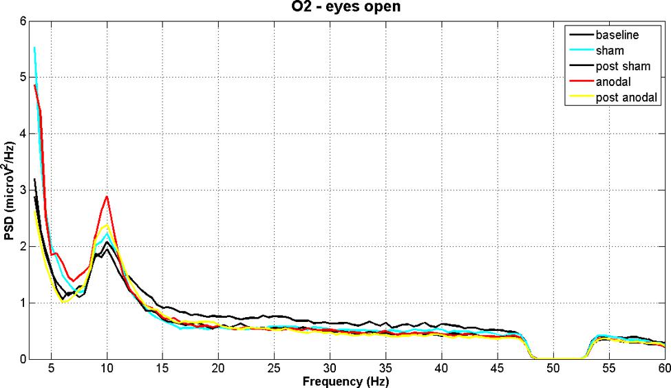
Eyes closed eyes open. Method used for spectrum analysis Fig. 15 17 18 Spectral analysis is based on the Fourier theorem which states that any waveform can be decomposed into a sum of sine waves at different frequencies with different amplitudes and different phase relationships.
Among various spectral analysis techniques we are focusing on Fast Fourier Transform FFT Wavelet Transform Event-Related SynchronizationDesynchronization ERSERD Complex Demodulation and Hilbert Transform. Max_frequency which represents maximum sampling frequency default value is 32. Spectral analysis is one of the standard methods used for quantification of the EEG.
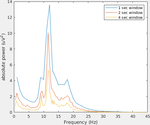
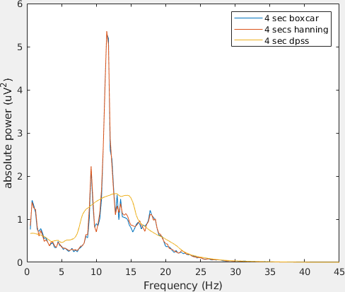
To calculate absolute and power spectrum as well as estimated and lowest frequencies for an EEG signal we will use the fft_eeg function. 51 Non-overlapping 4-sec epochs were weighted with a Hamming window and periodograms were then computed for these epochs using the Fast Fourier transform FFT. In this work PSD was calculated from EEG data during the processing of various visual stimuli.
Our analysis reporting is specific for these areas and also focused on three main region of interest ROIs. The final report was exported to be spectral_entropy_analysis_reportpdf. These include wavelet analysis and Fourier analysis with new focus on shared activity between rhythms including phase synchrony coherence phase lag and magnitude synchrony comodulationcorrelation and asymmetry.
The power spectrum indicates the power of each frequency component present in the source time domain waveform. Long to give about 50 sec playback time for each loop pass. Thirty-six subjects were randomized to either RT or a music comparison condition.
We conducted a controlled randomized study of the central nervous system effects of RT using spectral analysis of EEG activity. One of the most challenging predictive data analysis. Analysism and analysismlx for the experimental adjustment on different parameter settings of the spectral analysis.
No difference between normal controls and neurotic in. Normal oscillations in different frequency bands have an important role in cognitive processing in the frontal region. Four channels of EEG T4-T6 P4-02 T3-T5 P3-01 were recorded from several groups of control subjects and schizophrenia patients on analogue tape.
EEG spectra for each artifact-free 4-sec epoch were then aligned with 20-sec visually scored sleep. They were later digitized and analysed by computer. The mean normalized PSD for every EEG channel in the frequency ranges.
Spectrum Analysis of EEG Signals Using CNN to Model Patients Consciousness Level Based on Anesthesiologists Experience Abstract. Power spectra were computed for 30 second epochs of EEG per channel. The key issue is whether frequency oscillations of Electroencephalography EEG are related to cognitive task or not in occipital region.
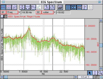
Spectral Analysis Use Acq Knowledge to obtain the power spectrum of the EEG. Spectral analysis is probably the most widely used computerized analysis of a digitized EEG. The acute central nervous system effects of relaxation techniques RT have not been systematically studied.
As a whole group and also when subdivided according to the type of delirium severity of cognitive decline or the type of central nervous system disease. First fast Fourier transform was computed and then squared to obtain the PSD estimate. The parameters of the function are.
Skip to Main Content. Spectral analysis of EEG was conducted for 51 elderly delirious patients meeting the Diagnostic and Statistical Manual of Mental Disorders III DSM-III criteria and for 19 controls. One of the most challenging predictive data analysis efforts is an accurate prediction of depth of anesthesia DOA indicators which has attracted growing atten.
The EEG spectral analysis is based on a set of frequency sub-bands. The power spectral density power spectrum reflects the frequency content of the signal or the distribution of signal power over frequency. Techniques used in digital signal analysis are extended to the analysis of electroencephalography EEG.
Researchers have mainly used wavelet transform WT 345678910111213141516 and time-frequency distributions TFD 17181920 to analyse the EEG spectral patterns. You will learn how to use them by seeing them applied to example data from resting state and visually evoked potential paradigms. Perform power spectral analysis on EEG data.
Monitoring Anesthetic Brain States The Spectral Analysis Of The Eeg Clinical Anesthesia Procedures
Time Frequency Analysis Of Eeg Data Fieldtrip Toolbox

Spectral Analysis Of Resting Electroencephalography The Multi Channel Download Scientific Diagram
Time Frequency And Spectral Analysis Fieldtrip Toolbox

The Power Spectral Density Of A Typical Eeg Channel With Superimposed Download Scientific Diagram

Eeg Still Breathing
Eeg Power Spectral Density Brainstorm
Monitoring Anesthetic Brain States The Spectral Analysis Of The Eeg Clinical Anesthesia Procedures

Eeg Power Spectral Density Brainstorm

Results Of The Frequency Tagging Analysis Power Spectrum Of The Eeg Download Scientific Diagram
Eeg Power Spectral Density Brainstorm
A More Negative Broad Band Spectral Exponent B Indexes A Steeper Download Scientific Diagram

Fronto Frontal Absolute Eeg Power Spectral Density V2 Hz In Wake Download Scientific Diagram

Spectral Analysis Of Intracranial Electroencephalogram Ieeg Top Download Scientific Diagram

Summary Of Eeg Data Analysis Pipeline Cross Spectral Density Between Download Scientific Diagram
Eeg Electroencephalography Spectral Analysis Research Biopac

The Power Spectral Density Plot Calculated From The Eeg Data Recorded Download Scientific Diagram

Sleep Eeg Multitaper Tutorial An Introduction To Spectral Analysis Part 1 Of 3 Youtube
Time Frequency And Spectral Analysis Fieldtrip Toolbox


Comments
Post a Comment